Utilizing 3D Models in Medical Teaching and Research: A Guide
Immersing into the dynamic sphere of medical teaching and research, one can immediately identify the essential role of visualization and hands-on learning. Emerging technologies such as 3D modeling have now created innovative platforms that transcend conventional methods of teaching anatomy, understanding complex pathologies, planning surgical procedures, and conducting valuable research endeavors. This guide explores the profound impact, potential, and application of 3D models in the medical realm.
Why 3D Models?
Two-dimensional diagrams, textbook illustrations, or even cadaveric dissections can offer valuable insights, but they often fall short in replicating the real-life complexities and intricacies found in human anatomy and disease processes. Cue 3D models – providing realistic, tangible, and patient-specific models that offer an unparalleled level of detail, precision, and customization.
Applications in Medical Teaching
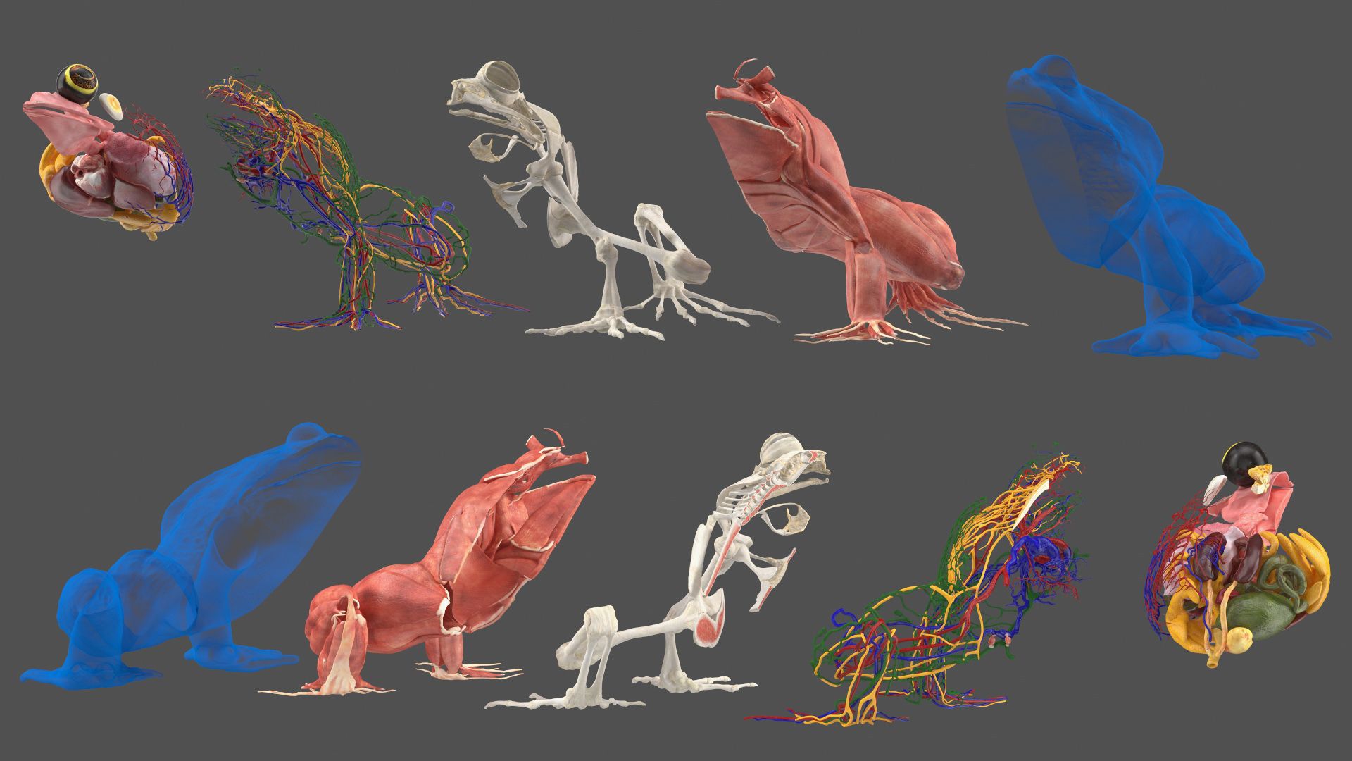
- Anatomical Education: 3D printed organ models accurately replicate anatomical structures, allowing students to see, touch, and understand the physicality and complexity of human anatomy, offering interactive and engaging learning experiences.
- Surgical Training: Intricate surgical procedures can be practiced on 3D models, offering surgical trainees a safer and more ethical platform for skill enhancement before performing on real patients.
Applications in Medical Research
- Device Testing and Innovation: 3D models are a valuable tool for testing new medical devices’ functionality, safety, and user compatibility.
- Personalized Medicine: Patient-specific 3D models ascertain personalized investigation and treatment methodologies for individualized patient care.
Implementing 3D Models in Medical Education and Research
- Decide on a Purpose: Identify what you aim to achieve with the 3D model. Be it teaching neuroanatomy, practicing surgical skills, or testing the latest cardiac device, this primary goal will guide the subsequent steps.
- Obtain Accurate and Detailed Data: The quality of the data significantly impacts the model. Clinical imaging techniques such as CT, MRI, or ultrasound can provide the necessary data.
- Use Appropriate Software: Various software tools allow you to transform clinical data into 3D printable models, from open-source ones like 3D Slicer, to advanced applications like Mimics.
- Choose the Right Printing Material: Your desired model’s purpose will dictate your selection. Flexible material can simulate soft tissues like blood vessels, while rigid material can depict bones or teeth.
- Test and Refine: Once your model is printed, evaluate it and refine it as necessary, ensuring it accurately mirrors what it’s intended to represent.
Cautions and Ethical Considerations
While 3D models introduce an array of valuable resources, potential ethical and legal considerations must be carefully navigated. This may include patient consent for the utilization of their medical data, respect for patient privacy/confidentiality, and consideration of cost implications.

Conclusion
The merger of 3D technology into the realms of medical education and research offers an exciting frontier for enhancing our understanding of the complex world of human health and disease. Whether it’s elevating the teaching experience, refining surgical skills, or empowering trailblazing medical innovations, 3D models are set to revolutionize the landscape of medical science. Embracing this technology today holds the promise of significantly augmenting the healthcare horizon of tomorrow.